Bowel pathology Radiology Cafe

MR enterography ( MRE ), also known as MRI small bowel study, is a non-invasive technique for the diagnosis of small bowel disorders. Note: This article is intended to outline some general principles of protocol design. The specifics will vary depending on MRI hardware and software, radiologist's and referrer's preference, institutional.
CT of Small Bowel GIST Tumors Imaging and Theory Part 2 YouTube

Ref: B-264/Imaging/MW/MRI of the small bowel v4. PDF: MRI of the small bowel [pdf] 181KB . YOU MUST ATTEND YOUR APPOINTMENT 1 HOUR PRIOR TO YOUR APPOINTMENT TIME . Failure to do so may mean your scan will be postponed. What is MRI (Magnetic Resonance Imaging) of the small bowel? This is a MRI examination of the small bowel and abdomen.
The Radiology Assistant Small Bowel Tumors
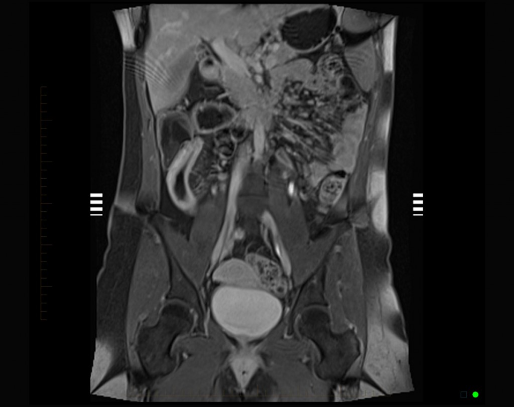
Imaging of the small bowel is most often performed on 1.5-T MRI, although the examinations can be performed on 3-T MRI with slight adaptation of the sequences. All sequences should be performed in breath-holds. The patient's position in the MRI can be supine or prone.
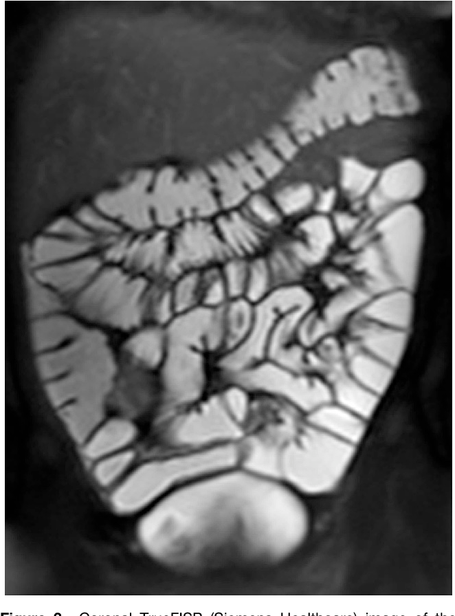
Representative small bowel MRI sequences. [A] TRUFIsequence small... Download Scientific Diagram

Small-bowel radiology has undergone dramatic changes in the past 2 decades. Despite important recent advances in small-bowel endoscopy, radiologic imaging remains important for patients suspected of having or with established small-bowel disease. Cross-sectional imaging techniques (computed tomography and magnetic resonance [MR] imaging), used to investigate both extraluminal abnormalities and.
MRI to evaluate small bowel Crohn's disease

MRI Small Bowel Imaging Page 5 of 8 order to provide a full and comprehensive report on your scan. A cannula (small needle) will be inserted into a vein in the arm or hand before the scan. The first is a muscle relaxant (Buscopan) used to settle bowel movement obscuring the area
MR Enterographic Manifestations of Small Bowel Crohn Disease RadioGraphics

MRI Small Bowel Study This leaflet explains about MRI Small bowel studies, including the benefits, risks and any alternatives and what you can expect when you come to hospital. If you have any further questions, please speak to a doctor or nurse caring for you. What is an MRI scan? Magnetic Resonance Imaging (MRI) is a type of scan that uses.
Small intestine in Crohn's disease, MRI Stock Image C029/4649 Science Photo Library

MRI of the gastrointestinal tract is gaining clinical acceptance and is increasingly used to evaluate patients with suspected small-bowel diseases. MRI may be performed with enterography or enteroclysis, both of which combine the advantages of cross-sectional imaging with those of conventional enteroclysis. In this paper, MRI features of primary small-bowel neoplasms, the most important signs.
Small intestine, MRI Stock Image C035/3600 Science Photo Library

Representatives from the Society of Abdominal Radiology Crohn's Disease-Focused Panel, the Society for Pediatric Radiology, the American Gastroenterological Association, and other international experts recently reported consensus recommendations for standardized nomenclature for the interpretation and reporting of CT enterography and MR enterography findings of small bowel Crohn disease.
MR Imaging of the Small Bowel Radiology

MRI Scanning for Small Bowel Disease Introduction This leaflet tells you about having an MRI scan for small bowel disease. The small bowel is difficult for doctors to get to with an endoscope (camera). It lies beyond the stomach and duodenum, and loops around for several metres to the right lower abdomen where it joins the large bowel.
Figure 3 from Small bowel imaging with MRI. Semantic Scholar
It's easy to find out more about treatment by giving us a call or completing our enquiry form. 01580 363158. Make an enquiry. Small bowel MRI or MR enterography uses an MRI scanner to produce detailed images of your small intestine to help identify small bowel conditions.
Small Bowel Obstruction due to Adhesions Small Bowel Case Studies CTisus CT Scanning

Cross-sectional imaging techniques are playing an increasing role in the evaluation of suspected small-bowel disorders, and a growing awareness of the risks of ionizing radiation exposure has prompted the exploration of alternative imaging techniques. Advantages of magnetic resonance (MR) imaging include a lack of ionizing radiation, the ability to provide dynamic information regarding bowel.
MRI to evaluate small bowel Crohn's disease
Small bowel cancers are difficult to diagnose. For this reason, people suspected of having small bowel cancer often need multiple tests and procedures to locate the cancer or rule out a cancer.. They can show the location and size of small bowel cancer. Tests might include MRI, CT and positron emission tomography, also called a PET scan.
Small Bowel Crohn Disease at CT and MR Enterography Imaging Atlas and Glossary of Terms

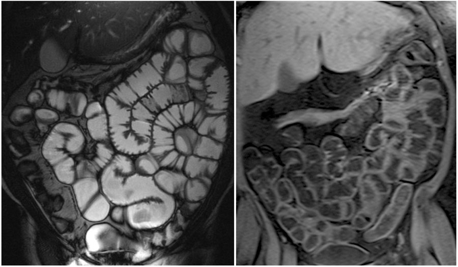
The purpose of utilizing an oral contrast agent is to achieve specific signal intensities in the bowel lumen on different types of MRI images. On T2-weighted images, the aim is to produce a high signal intensity in the bowel lumen, while on T1-weighted images, a low signal intensity in the lumen is desired.
Bowel pathology Radiology Cafe

During your MRI scan. You will be asked to drink between 800 to 1500mls of the Mannitol sugar solution after you have had safety checks with a radiographer or radiology assistant. This solution should reach the end of your small bowel within 40 to 60 minutes. Sometimes this can take longer. The MRI scan can then begin.
Small bowel MRI enteroclysis or follow through Which is optimal?
MRI techniques available to evaluate the small bowel include MR enterography (MRE) and MR enteroclysis. In MR enteroclysis, enteric contrast is administered directly via a nasoenteric tube, providing superior distention of small-bowel loops compared to oral ingestion in MR enterography. However, MR enteroclysis remains of limited availability.
Small bowel lymphoma Radiology at St. Vincent's University Hospital
the body. The MRI scanner does not use X-rays. This is a scan to look at your small bowel. It may help your doctor diagnose inflammation, blockages and other problems. How long will it take? First you will be given a drink to have over 1 hour. The MRI scan usually takes between 30 and 45 minutes. The time will vary for each patient.
.