Coronal section through the brain Diagram Quizlet

The Brain Biodiversity Bank at Michigan State University. Home >> Atlases >> The Human Brain Atlas >> Coronal Montage of the Human Brain. Human Brain Menu. Coronal Montage. Horizontal Montage. Sagittal Montage.
Coronal sections of the brain Anatomy Kenhub
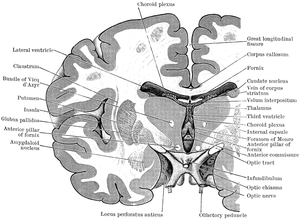
Show details. Coronal section of brain through anterior commissure, Caudate nucleus, Internal Capsule, Putamen, Globus pallidus, Claustrum, Insula, Optic Chiasma, Third Ventricle, Anterior Commissure, Columns of fornix, Cavity of septum pellucidum, ANterior Cornu, Corpus callosum Henry Vandyke Carter, Public Domain, via Wikimedia Commons.
Coronal section of human brain plastinated specimen with 5 parts, plastinated human specimen
Coronal sections through the brain. fig5.8 fig5.9 fig5.10 fig5.11 fig5.12. The five coronal sections through the brain shown in (Figures 5.8, 5.9, 5.10, 5.11, and 5.12), were taken from Sylvius4 and should resemble the brains that are available for examination in the laboratory. Remember, areas with little or no myelin appear dark and are.
Coronal section through brain Diagram Quizlet

Home >> Atlases >> The Human Brain Atlas >> External Lateral View of the Human Brain. Coronal Sections of the Human Brain. Click on a line to see a coronal section at that level. All images on this site are copyrighted and produced with the support of public funds.
Coronal section of a healthy brain showing normal anatomy of basal baglia Stock Illustration

Figure 1. These illustrations show the (a) coronal and (b) sagittal sections of the human brain. In other surgeries to treat severe epilepsy, the corpus callosum is cut instead of removing an entire hemisphere. This causes a condition called split-brain, which gives insights into unique functions of the two hemispheres.
Posterior Cerebral Artery
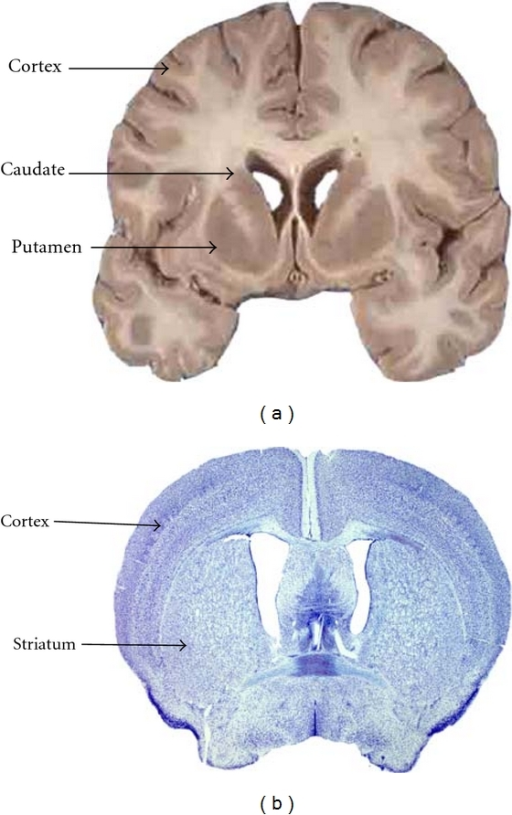
Coronal sections of the brain allow deep tissue structures to be visible. A cut through the anterior portion of the temporal lobe shows the amygdala, a region important for emotion, located in the medial temporal lobe. The regions of the basal ganglia are also visible; the striatum, which consists of the caudate and the putamen, and the globus.
Coronal level 2060 as Cell Stain

Figure 12.3.3 12.3. 3: Coronal Section of the Brain. The coronal section shows the layers of the cerebrum. The superficial gray matter is called cerebral cortex, while the deep gray matter is organized in basal nuclei. The superficial and deep regions are connected by myelinated axons (white matter) called tracts.
(a) Coronal section of a human brain showing the cortex Openi
(C) Coronal section image from a Nissl-stained brain at the approximate center of the IPN. Nissl reveals putative IPN subdivisions as regions containing neurons of different sizes and densities. (D-F) Coronal section images of the IPN from three brains immunohistochemically stained with a pair of antibodies as indicated.
Posterior Cerebral Artery
The coronal suture runs from side to side across the skull, within the coronal plane of section (see Figure 7.3.3). It joins the frontal bone to the right and left parietal bones.. The brain case is that portion of the skull that surrounds and protects the brain. It is subdivided into the rounded top of the skull, called the calvaria, and.
27 Brain Sections Neupsy Key

Atlas of the Brain in Coronal Section. Atlas of Gross Topography of the Brain - Swenson. Go to main atlas index.
Coronal Section Through the Cerebrum ClipArt ETC
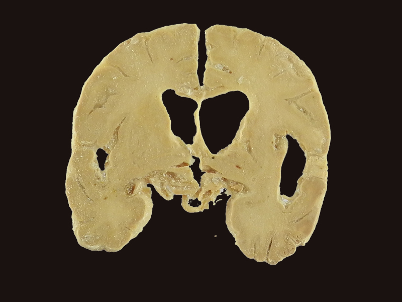
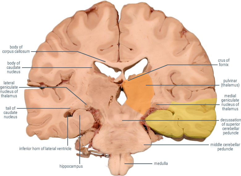
Coronal section of the brain at the level of the thalamus. The frontal and temporal lobes are observed in their previously described locations. The body of the corpus callosum forms the roof of the body of the left and right lateral ventricles, which are separated from each other by the septum pellucidum. The insula of Reil and the Sylvian.
coronal section of brain Diagram Quizlet

Coronal Section View. Coronal sections of the brain allow deep tissue structures to be visible. A cut through the anterior portion of the temporal lobe shows the amygdala, a region important for emotion, located in the medial temporal lobe. The regions of the basal ganglia are also visible: the striatum (which consists of the caudate and the.
Coronal Sections of the Cerebral Hemispheres ClipArt ETC
We generated a whole-brain molecular atlas by capturing the spatial patterns of gene expression in the adult mouse brain using ST ().We hybridized 75 coronal sections from one brain hemisphere that covered the entire AP axis onto ST arrays (Fig. 1A and fig. S1).Using a computational framework designed to generate reference maps (), we aligned each imaged brain hemisphere section, including the.
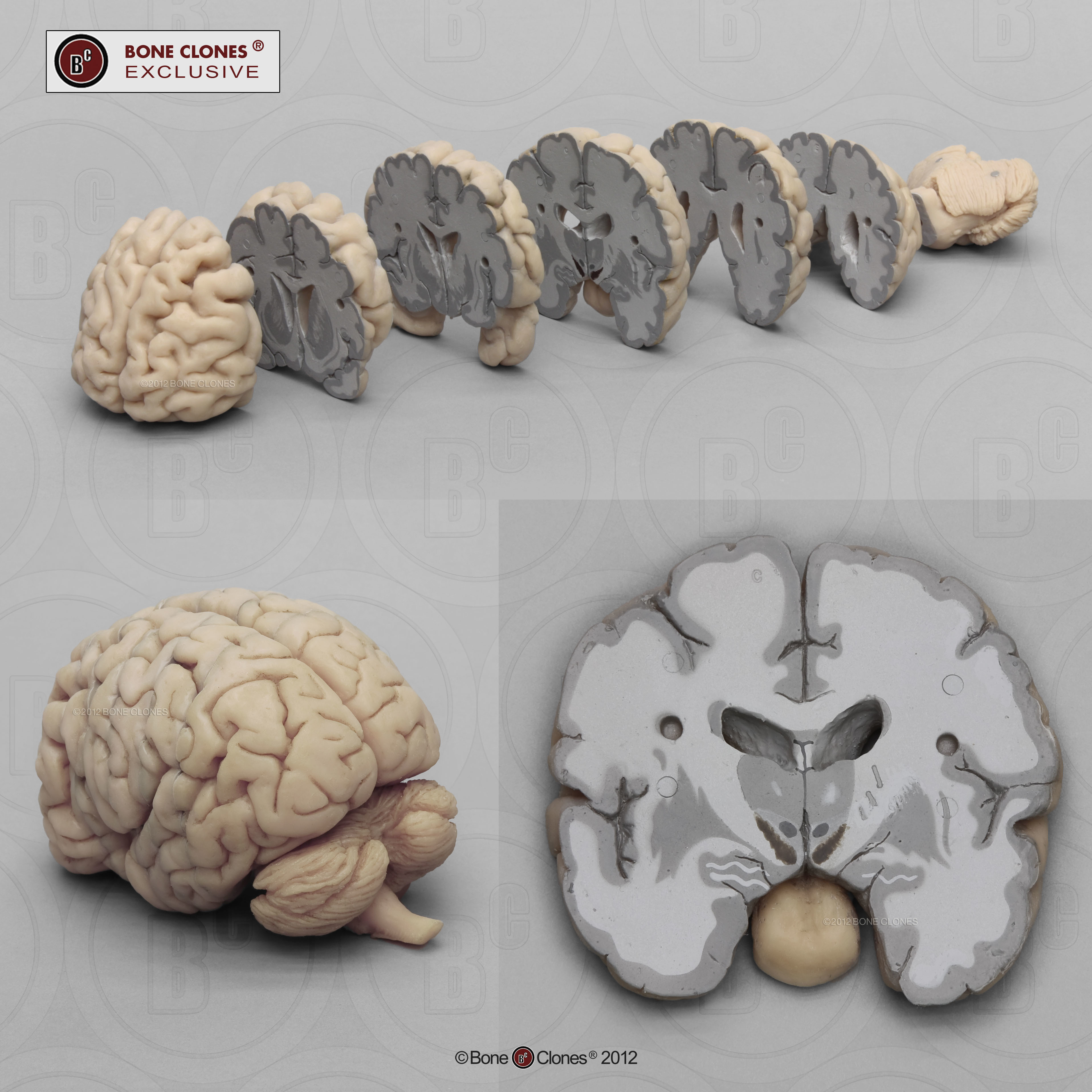
Human Brain Multiple Coronal Sections Bone Clones, Inc. Osteological Reproductions
Functional Neuroanatomy. Home
27 Brain Sections Neupsy Key

Figure 1: Lobes of the cerebrum, lateral view, with inset showing insular lobe deep to lateral fissure. Figure 2: Brodmann areas of the cerebral cortex, A. Lateral view and B. Medial view. Figure 3: Gyri and sulci of the lateral surface of the cerebrum. Figure 4: Gyri and sulci of the medial surface of the cerebrum.
The rostral surface of a coronal section of brain through the level of the anterior tubercle of

The frontal or coronal plane is a vertical plane in a medial to lateral direction, dividing objects into front and back pieces. The sagittal plane is also a vertical plane but in a rostral-caudal direction, meaning it divides objects into right and left regions.. Gray matter is primarily neuronal cell bodies and dendrites. In the brain, the.
.